Content
- What is histology
- What is histology in gynecology
- Histology of the stomach
- What is histology in oncology?
- Histology price
- Reviews
The world of living beings has been interested in scientists from all over the world for many decades. Moreover, numerous laboratory studies have taken a significant step in a given direction, facilitated the fate of mankind. Histology speeds up diagnosis, helps to prescribe adequate treatment.
What is histology
This is the science of tissues, which allows timely detection of a progressive pathological process even at the cellular level. A thorough analysis of this biological material reveals cancer cells, structural mutations through microscopy. Using special equipment, foreign bodies and their detailed characteristics are determined with maximum accuracy. This is especially important in the light of the upcoming study, as the patient’s chances of full recovery only increase.
What histology studies
Tissues are those important body structures that begin the functionality of an organic resource. Answering the main question about what is the object of study in the field of histology, it is necessary to focus on this invisible simple philistine biological material. For scientists, tissue is a storehouse of useful information by which to judge the viability of the organism as a whole, its weak points, and future pathologies. The prevailing diagnosis will be made accurately, and the disease can be treated with drugs (conservatively) at an early stage..

What histology shows
This science is engaged in the microscopic study of intracellular structures. The main areas are five types of cells, among them epithelial, nervous, connective muscle tissue, blood. The results of histology help determine the presence of a pathological process and make a final diagnosis. In gynecology, this is a real chance to determine oncological diseases and the causes of pathological pregnancy. With a timely response to the problem, the woman is waiting for surgical measures, curettage with a favorable clinical outcome.
If histology is interested, what is it, a specialist will tell. He will tell you what this invasive laboratory study shows. So, from the interpretation of the analysis in histology, you can determine:
- inflammatory process;
- impaired systemic circulation;
- the fact of internal hemorrhage, the presence of thrombosis;
- the presence of cancer cells;
- the presence of malignant neoplasms and their parameters;
- metastases of neighboring organs.
Histology Analysis
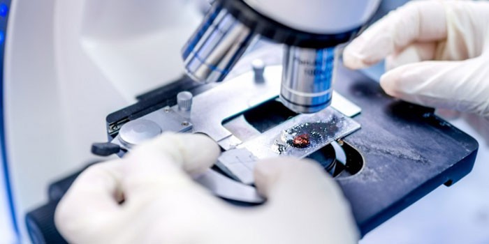
Laboratory research can be done exclusively in a hospital with modern equipment, as well as a biopsy. In modern medicine, this is a reliable diagnostic method that determines pathology even at the cellular level. Histological analysis examines the biological material, which is the particles of the epithelial layer of the internal organs, systems. It is carried out simultaneously with a biopsy, which just involves taking a bunch of living cells for further research.
What is histology in gynecology
Such a procedure is often carried out in modern gynecology, is a reliable method for the diagnosis of extensive pathologies of the uterus and its appendages, and timely identifies inflammatory and infectious processes of the cervix. Women who are faced with the problem of a missed pregnancy or miscarriage in the early stages, are well aware of what histology is in gynecology. This laboratory study helps determine the cause of the reproductive system pathology..
Uterine histology
This morphological analysis determines the structure of the cells, so it immediately notices their mutations in the presence of cancer. In order for the histology of the endometrium to help determine the final diagnosis, the doctor insists on carrying out preparatory measures. An integrated approach to the problem increases the informational content of laboratory tests, helps to more likely begin intensive therapy with drugs. Here are the prerequisites before going to a biopsy:
- For two weeks, exclude from the diet food additives that happened to be bought in an online store or pharmacy.
- For 3-5 days, refrain from sexual intercourse, strengthen compliance with intimate genital hygiene.
- Be sure to perform a general blood test, a study for the presence of genital infections, a bacteriological smear.
- A laboratory study should be carried out before planned menstruation, another period of the menstrual cycle for diagnosis is excluded.
- It is important to pre-arrange any medication with a specialist, since bleeding can be triggered during a laboratory test.

Histology after a frozen pregnancy
If the fetus died in the second trimester, the doctor performs an urgent curettage of the uterine cavity, followed by histological examination. It is extremely important to timely determine the cause of a frozen pregnancy, to prevent its recurrence. If properly treated, there is every chance to experience the joy of motherhood. Histology in an aborted pregnancy examines the tissues of a dead embryo to determine in conclusion the causes of miscarriage. It:
- viral and infectious processes;
- hormonal imbalance of the female body;
- diabetes;
- genital infections
- uterine anomalies.
Histology after curettage
The study itself involves the removal of part of the uterine epithelium. They perform an operation before menstruation in order to reduce the amount of blood loss and speed up the process of regeneration of damaged tissues. Biological material is taken for histological analysis after curettage. First of all, it is placed in a special solution to prevent cell decay. Then treated with paraffin and already in the hardened form perform a small section. Then they are stained in color, examined under a microscope. In this way, healthy cells can be distinguished from cancerous modifications..
Histology of the stomach
If the doctor suspects malignant tumors of the digestive tract, the patient will have to perform histology of the stomach, for example. The resulting decryption gives an idea not only about the presence of cancer, but also directly about the tumor itself. The histology of the stomach determines the size of the focus of the pathology, the cellular composition, the presence of metastases. This is an informative study, so doctors perceive the positive answer about the presence of cancer as the final diagnosis. In addition to histology, hysteroscopy may be required to clarify.

What is histology in oncology?
Before ordering such a laboratory study at a tangible price, you need to understand whether it is required to be carried out in a specific clinical picture. If this is a suspicion of malignant tumors, the answer is unequivocally affirmative. Cytology and histology are the basis of a comprehensive diagnosis, since such studies reveal cancer cells at an early stage of their formation. Decryption helps to quickly begin treatment, provide a stable therapeutic effect.
Histology price
All patients are interested in how much histology costs. The cost of the study depends on the alleged focus of the pathology, the city of the patient, the clinic and the reputation of the specialist who conducts this laboratory study. The difference is not always noticeable, therefore it is better to rely not on the criterion of “inexpensive”, but on the professionalism of a specialist.
Reviews
Marina, 34 years old
I read the catalog with the research prices of one metropolitan laboratory. I must say right away that the price of histology is adequate, affordable. The difference with other analyzes is palpable. I went to the laboratory with my problem on the recommendation of the attending physician. I had an enlarged mole – I had to check. The sensations are not pleasant, the skin is still frost.
Anna, 31 years old
Unlike others, my problem was not so global. I needed histology before cauterizing erosion. I read the description of this study, but in reality everything is worse. This is painful as a piece of living flesh is plucked off. Decoding of histology showed that there are no problems. I’m glad I checked, but the memories are not very.
Olesya, 26 years old
I went through a study at a histopathological center before cauterizing erosion. Cells are nipped off, then part is placed on glass, and part is placed in a container with liquid. Sensations – pain, then easier. In the evening it hurt in the lower abdomen, I had to take painkillers, a couple of days there was a discharge. Affordable price.
Similar articles
- CTG during pregnancy – what it is and how it is done. What is shown by cardiotocography of the fetus and interpretation of the results
- Analysis of urine for white blood cells is the norm for children and adults. What do elevated white blood cells mean in a urinalysis
- Ultrasound of the abdominal cavity – preparation for the study. What to eat and drink before an ultrasound of the abdomen